


Fundus Flourescein
Angiography
What Is Fundus Flourescein Angiography
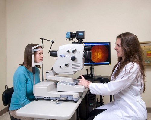
Fundus Fluorescein Angiography are tests that use special cameras to photograph the structures in the back of the eye. These tests are very useful for locating the damage to the blood vessels that nourish the retina (light sensitive tissue) and in turn, checking on the health of the retina itself. In both tests, a colored dye is injected into a vein in the arm of the patient. The dye travels through the circulatory system and reaches the vessels in the retina and those of a deeper tissue layer called the choroids. Neither of the tests uses any harmful forms of radiation.
Fluorescein is a yellow dye, which glows in visible light, it requires a special digital camera sensitive to these light rays This angiography helps your doctor make the correct diagnosis and plan the best course of treatment especially in diseases like age related macular degeneration (AMD).
How Is Fundus Angiography Done
Before the procedure, you will be asked questions about your general health and the medications that you are using. A self-explanatory consent form, which explains the side effects in detail, will be provided to you. You will have to give your consent before the procedure. You should have a light meal before undergoing the procedure and must be accompanied by a family member or friend.
What Are The Side-Effects
Fluorescein angiography are considered very safe and serious side effects from these tests are uncommon. However, there is the possibility that a patient may have a reaction to the dyes. While fluorescein contains no iodine and is safe in patients known to be allergic, Some people may experience slight nausea after the dye injection, but the feeling usually passes quickly. Patients who are allergic to the dye can develop itching and a skin rash. These symptoms generally respond quickly to oral medications such as anti-histamines or steroids.
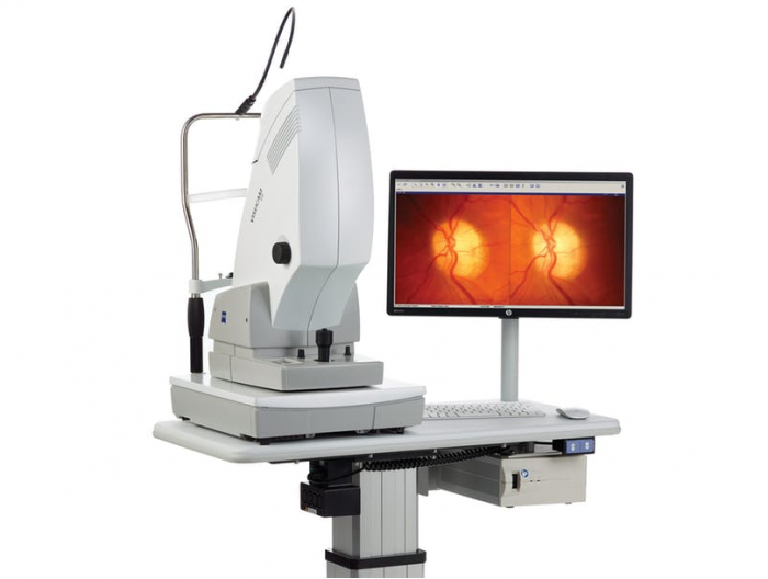
- CARL ZEISS VISUCAM PRO NM/FA The Zeiss VISUCAM PRO NM Non-Mydriatic Fundus Camera increases the quality and simplicity of fundus imaging.
- Red-free
- Color Fundus
- Anterior Segment
- Fluorescein angiography
- Blue and red wavelength range
- Fast Windows XP computer with database
- Quick image transfer via network, USB stick or DVD/CD
- ZEISS Telecentric Optics with integrated, medical grade digital sensor
- Network ready and with convenient image transfer via USB device, or DVD

Preparation for Fundus Fluorescein Angiography
Before the FFA procedure, patients are typically asked to avoid driving or operating heavy machinery for a few hours, as the dye used can temporarily affect vision. Patients may also be advised to avoid certain medications that could interfere with the dye’s effects. The hospital staff will provide detailed instructions on how to prepare for the procedure.
During the FFA procedure, a small amount of fluorescein dye is injected into a vein, usually in the arm. The dye then circulates through the bloodstream and reaches the blood vessels in the eye. A specialized camera is used to take a series of photographs as the dye passes through the eye’s blood vessels. This allows the eye doctor to assess the health and function of the retina and other structures.
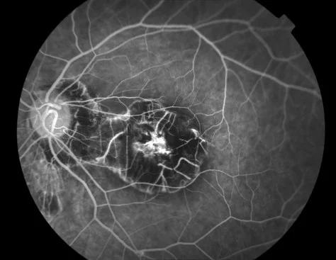
The images obtained from the FFA procedure are carefully analyzed by our experienced retina specialists. They look for any abnormalities in the blood vessel patterns, leaks, or other signs that may indicate underlying eye conditions, such as diabetic retinopathy, age-related macular degeneration, or retinal vascular occlusions. The results of the FFA are then used to develop a personalized treatment plan for the patient.
To schedule an appointment or learn more about our services, please don’t hesitate, Hurry Contact us and Schedule an appointment.
FAQs About Fundus Fluorescein Angiography
Fundus Fluorescein Angiography is a diagnostic procedure that allows eye doctors to visualize the blood vessels in the back of the eye, helping them to identify and manage various retinal and vascular conditions.
Fundus Fluorescein Angiography is generally a safe procedure, but there is a small risk of allergic reaction to the dye. Our medical team closely monitors patients during and after the procedure to ensure their safety.
The FFA procedure typically takes 15-30 minutes, depending on the complexity of the case and the number of photographs required.
FFA involves minimal discomfort, only a brief needle stick for injection. Eye photography is completely painless throughout.
Total time including preparation, testing, and monitoring is typically 45-60 minutes from arrival to departure completion.
Serious reactions are extremely rare. Most people experience no problems beyond temporary yellow urine and skin tinge.
Dilated pupils impair vision temporarily. Arrange transportation or wait several hours until pupils return to normal size.
Slight yellow tinge may occur temporarily but fades within hours as dye is eliminated through kidneys.
Frequency depends on your condition. Some patients need annual testing while others require monitoring every few months.
Most insurance plans cover medically necessary FFA testing. We verify coverage before scheduling your examination appointment.
FFA is generally avoided during pregnancy unless benefits clearly outweigh potential risks in urgent situations carefully.
Fluorescein dye contains no iodine. Iodine allergies don't prevent FFA testing safely in most patients successfully.
No, FFA causes no eye damage. The bright camera flashes create temporary light sensitivity without harming structures.
Yes, children can safely undergo FFA when medically necessary with appropriate preparation and gentle pediatric approaches.
FFA helps diagnose diabetic retinopathy, macular degeneration, vascular occlusions, inflammatory diseases, and many other retinal conditions.