


Introduction:
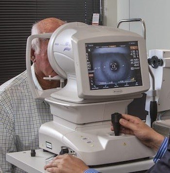
Optical Coherence Tomography (OCT) has transformed the field of ophthalmology, allowing eye specialists to examine the retina in exceptional detail. This advanced imaging technique is instrumental in diagnosing numerous eye conditions, especially those affecting the retina and optic nerve. However, as powerful as OCT is, it’s not without its limitations. One such limitation is the phenomenon known as “Green Disease.” This term refers to a specific challenge in interpreting OCT images accurately, where color-coded “green” areas on the scan may mistakenly suggest healthy tissue when, in fact, early signs of disease are present. Understanding Green Disease is essential for both patients and eye care providers to ensure accurate diagnosis and prevent potentially serious eye conditions from going undetected. In this article, we’ll explore what Green Disease in OCT is, why it happens, and how to address it for the most accurate eye health assessments.
Why Does Green Disease Occur in OCT Scans?
Green Disease is a consequence of the inherent limitations of OCT technology and the human anatomical variations that impact scan interpretation. OCT works by capturing cross-sectional images of the retina and mapping these areas into color-coded zones. The algorithms behind OCT imaging rely on preset thresholds to classify tissue as healthy or unhealthy. However, these thresholds are general averages and do not account for the nuanced variations that exist from one person to another.
For instance, people naturally have differences in the thickness of their retinal nerve fiber layers or in the structure of their optic nerve. These differences mean that a “normal” OCT scan for one person may not look the same for another, even if both are healthy. Consequently, the color-coded maps may fail to capture subtle changes or early disease signs, especially in patients with atypical anatomy. Furthermore, OCT scans are sometimes used as a standalone diagnostic tool. While OCT is incredibly useful, relying solely on OCT images without additional assessments can increase the risk of Green Disease, as it does not offer the full picture of a patient’s retinal health.
Conditions Commonly Misdiagnosed Due to Green Disease
Green Disease poses a particular risk in the diagnosis of certain eye conditions. One of the most significant is glaucoma, where early stages often involve subtle thinning of the retinal nerve fiber layer that may not immediately appear on an OCT scan. Since glaucoma can progress without obvious symptoms, it’s easy to miss in its early stages if clinicians are relying on OCT’s “green” areas as definitive proof of health.
How to Address Green Disease in OCT Interpretation
To mitigate the risk of Green Disease and ensure a comprehensive assessment, eye care professionals can employ a variety of strategies. One effective approach is to use OCT in conjunction with other diagnostic tools. For example, visual field tests can provide valuable information about the functional aspects of a patient’s vision, which may reveal early signs of conditions like glaucoma that an OCT scan alone might miss. Additionally, fundus photography and angiography are complementary imaging techniques that offer different views of retinal health, allowing practitioners to detect abnormalities that could be hidden in OCT’s green areas.
Conclusion
Green Disease is a reminder of the complexities involved in diagnosing eye health conditions and the limitations of relying solely on OCT imaging. While OCT is an invaluable tool, its interpretations are not foolproof, and a green-coded area should not be considered a guarantee of perfect retinal health. A holistic, multi-modal approach to eye exams, guided by experienced professionals, ensures that even the subtlest signs of disease are not overlooked. At Retina Specialty Hospital, we are committed to providing precise, comprehensive care, using OCT alongside other diagnostic methods to deliver clarity and accuracy for our patients. If you’re experiencing vision changes or have questions about your OCT results, contact us today to schedule a consultation and explore the full scope of your eye health.