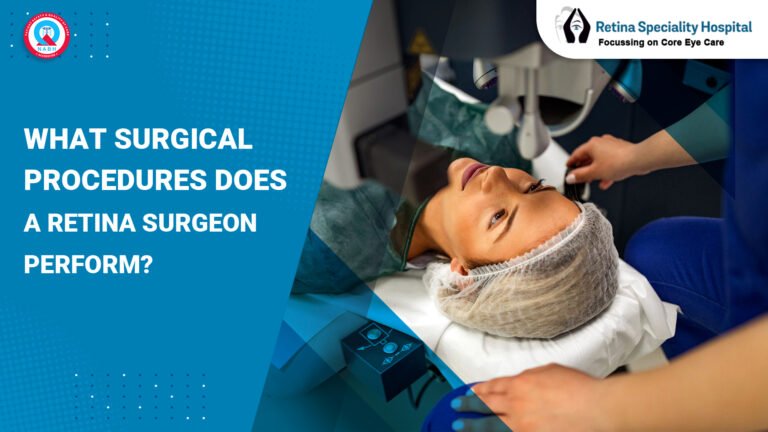


Glaucoma is called the silent thief of sight for a very specific reason. According to the World Glaucoma Association, over 80 million people worldwide are affected by glaucoma and nearly half of them do not know they have it. The OCT RNFL test is one of the most powerful tools available today for detecting glaucoma damage before any vision loss is felt by the patient. Patients who complete their RNFL test in Indore at our clinic regularly ask the same question: what happens next? This guide explains every step after the scan, from how your doctor reads the report to what abnormal results mean for your treatment plan, helping you feel confident and informed throughout the entire process.
Understanding your RNFL OCT scan in Indore results is not complicated when explained clearly. Knowledge of the process helps you work more effectively with your specialist toward protecting your vision long term.
How Is the OCT RNFL Test Done? A Guide for First-Time Patients
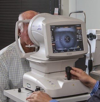
The OCT RNFL test uses optical coherence tomography technology to measure the thickness of the retinal nerve fiber layer surrounding the optic nerve head. This layer contains the axons of the ganglion cells that carry visual information from the retina to the brain.
During the scan, you sit in front of the OCT machine and rest your chin on a support. The machine directs a safe, painless beam of near-infrared light into your eye. The entire scan takes less than 5 minutes per eye and requires no injections, no contact, and no dilation in most cases.
The machine captures cross-sectional images of the nerve fiber layer and measures thickness in microns across four quadrants: superior, inferior, nasal, and temporal. This data is compared against a normative database of thousands of age-matched healthy eyes to identify any thinning that may indicate early nerve damage.
When Will I Get My OCT RNFL Test Report?
At the best eye hospital for RNFL scan in Indore, most patients receive their printed and digital OCT report on the same day as their scan. The machine generates a detailed printout immediately after the scan is completed.
However, the printed data alone does not constitute a diagnosis. A qualified eye specialist review after OCT is required to interpret the measurements correctly in the context of your age, eye pressure, family history, and any existing symptoms you may have reported.
Your doctor will typically review the report with you during a same-day or next-day consultation appointment. At our clinic, every RNFL OCT scan in Indore is reviewed by a glaucoma-trained specialist before results are communicated to the patient with full explanation.
How Are OCT RNFL Test Results Interpreted?
Interpretation of RNFL thickness reports is a structured clinical process that goes far beyond simply reading numbers on a page. Your specialist looks at multiple layers of information in your report simultaneously.
The OCT printout displays a color-coded map of your nerve fiber layer. Green areas indicate thickness within the normal range for your age group. Yellow areas indicate borderline thinning that requires monitoring. Red areas indicate significant thinning that is outside normal limits and may represent glaucoma damage or early nerve degeneration.
Your doctor also examines the deviation map, the TSNIT curve (temporal, superior, nasal, inferior, temporal), and the global average RNFL thickness value. All of these data points together build a complete picture of your optic nerve fiber layer assessment and current eye health status.
What Is Normal RNFL Thickness?
Normal RNFL thickness in a healthy adult eye typically falls between 80 and 110 micrometers as a global average. However, normal values vary by age, as the nerve fiber layer naturally thins very slightly with age in all individuals throughout life.
The four quadrants have different normal thickness ranges. The superior and inferior quadrants are naturally thicker, typically between 100 and 130 microns, as they carry more nerve fibers. The nasal and temporal quadrants are thinner by comparison, typically between 60 and 80 microns in healthy eyes.
Your RNFL test in Indore report will indicate whether each quadrant falls within the normal, borderline, or outside normal limits category based on your specific age group comparison. A single value outside normal range does not automatically confirm glaucoma and must be evaluated in full clinical context by your doctor.
What Happens If OCT RNFL Results Are Abnormal?
Abnormal results on an RNFL OCT scan in Indore do not automatically mean you have glaucoma. Several conditions beyond glaucoma can cause RNFL thinning, including optic neuritis, previous retinal vein occlusion, myopia, and certain neurological conditions.
When results show red or yellow zones, your eye specialist review after OCT will include the following next steps to clarify the diagnosis accurately.
- Visual field test (perimeter) – Maps your peripheral vision to detect any functional loss corresponding to the structural damage seen on OCT.
- Intraocular pressure measurement – Elevated eye pressure is a major risk factor for glaucoma and is assessed alongside RNFL data.
- Optic nerve head photographs – High-resolution images of the optic disc help confirm whether thinning patterns match glaucoma-specific structural changes.
- Corneal thickness measurement (pachymetry) – Affects accurate pressure readings and influences overall glaucoma risk assessment.
- Gonioscopy – Examines the drainage angle of the eye to determine whether open-angle or angle-closure glaucoma is present.
These additional tests together with your OCT RNFL test results allow your specialist to make an accurate diagnosis and recommend the correct treatment plan for your specific situation.
| Result Color | RNFL Status | Clinical Meaning | Next Step |
| Green | Within normal limits | No significant thinning detected | Routine annual monitoring |
| Yellow | Borderline thinning | Possible early changes present | Repeat OCT in 6 months |
| Red | Outside normal limits | Significant thinning detected | Full glaucoma workup immediately |
| Asymmetry flag | Difference between eyes | May indicate early unilateral damage | Detailed optic nerve review |
| Inferior thinning | Early glaucoma pattern | Most common initial glaucoma sign | Visual field test same week |
| Superior thinning | Advanced or focal damage | Associated with inferior visual field loss | Urgent specialist review |
| Global average low | Generalised thinning | May indicate advanced nerve damage | Immediate treatment planning |
Do I Need Follow-Up Tests After the OCT RNFL Scan?
Yes, follow-up testing is an essential part of the early glaucoma detection process and should not be skipped even when initial results appear normal. A single OCT scan captures one point in time and cannot reveal whether thinning is progressive or stable.
Your specialist at the best eye hospital for RNFL scan in Indore will recommend a follow-up schedule based on your individual risk profile. Patients with normal results and no risk factors typically return annually. Patients with borderline or abnormal results may need follow-up every 3 to 6 months to track any progression accurately.
Can OCT RNFL Results Change Over Time?
Yes, OCT RNFL test results can and do change over time. In patients with glaucoma, progressive thinning of the nerve fiber layer is the clearest indicator that the disease is advancing despite treatment.
Serial OCT scans allow your doctor to generate a progression analysis report that compares your current scan with all previous scans performed at the same clinic. This trend analysis is far more clinically valuable than any single scan result alone because it shows the rate of change over months and years.
This is why staying with the same RNFL OCT scan in Indore facility matters. Consistent machine calibration and scan protocols make trend comparisons more accurate and clinically meaningful for your long-term care.
What Happens If Results Are Abnormal After the OCT RNFL Test?
When RNFL thickness analysis confirms abnormal results, your specialist will develop a structured treatment plan tailored to your diagnosis and severity level. The approach depends on whether glaucoma is confirmed or whether findings are classified as glaucoma suspect.
For confirmed glaucoma patients, treatment options discussed at our eye treatment in Indore clinic include the following interventions.
- Prescription eye drops – Prostaglandin analogues or beta blockers to lower intraocular pressure and slow nerve damage progression.
- Laser therapy (SLT) – Selective laser trabeculoplasty improves fluid drainage from the eye without incisions.
- Minimally invasive glaucoma surgery (MIGS) – For moderate cases where drops alone are insufficient to control pressure.
- Traditional glaucoma surgery (trabeculectomy) – Reserved for advanced cases with significant uncontrolled pressure and nerve damage.
For glaucoma suspect patients, treatment focuses on close monitoring with repeat RNFL test in Indore scans every 4 to 6 months and lifestyle modifications to reduce risk factors.
Is Repeat OCT RNFL Scan Necessary?
Yes, repeat RNFL OCT scan in Indore is not just necessary but essential for effective glaucoma management. Glaucoma is a chronic progressive condition and a single normal result does not guarantee that damage will not develop in the future.
Repeat scans serve three important clinical purposes. First, they confirm whether a previously abnormal result represents true pathology or a scan artifact caused by patient movement or poor signal quality. Second, they track progression rate in diagnosed patients to evaluate treatment effectiveness. Third, they provide reassurance and a documented baseline for patients in high-risk categories such as family history, high myopia, or elevated eye pressure.
Our best eye hospital for RNFL scan in Indore retains all previous scan data in a secure digital system so that every follow-up scan can be compared against your complete scan history accurately.
How Often Should OCT RNFL Be Done?
The recommended frequency of OCT RNFL test scans depends on your individual diagnosis, risk level, and treatment status. Here are the evidence-based guidelines followed by our specialists.
- Healthy adults with no risk factors – Every 1 to 2 years as part of a routine comprehensive eye examination.
- Glaucoma suspects – Every 6 months for the first 2 years to establish a reliable baseline trend.
- Confirmed glaucoma patients on treatment – Every 4 to 6 months to monitor treatment response and progression rate.
- Post glaucoma surgery patients – Every 3 months in the first year following the procedure.
- Patients with family history of glaucoma – Every year from the age of 35 onwards regardless of current symptoms.
Consistent monitoring through serial RNFL tests in Indore scans remains the gold standard for protecting vision in glaucoma patients worldwide according to the World Glaucoma Association guidelines.
CASE STUDY:
A 52-year-old retired school principal in Indore came to our clinic for a routine eye check-up with no specific complaints. He had no visual symptoms, normal eye pressure readings at 16 mmHg, and no family history of glaucoma that he was aware of. His first RNFL OCT scan in Indore showed borderline inferior thinning in the left eye flagged as yellow on the color-coded report.
Our glaucoma specialist recommended a 6-month follow-up scan rather than immediate treatment, as the findings were borderline and could represent normal variation. At 6 months, the inferior thinning had progressed slightly and a visual field test confirmed a small but reproducible superior arcuate scotoma corresponding to the area of thinning detected on OCT.
A diagnosis of early primary open-angle glaucoma was confirmed. The patient was started on once-daily prostaglandin eye drops and his next RNFL test in Indore scan at 12 months showed stabilization with no further progression. He continues 6-monthly monitoring at our clinic and his vision has remained fully preserved through early detection enabled by the OCT RNFL test process.
FAQ’s About OCT RNFL test
Q1. What happens after OCT RNFL test results are received?
Your specialist reviews RNFL thickness data and recommends follow-up tests, monitoring schedules, or treatment accordingly.
Q2. How do doctors analyze RNFL thickness after OCT scan?
Doctors compare your RNFL thickness against age-matched norms using color-coded maps, global averages, and quadrant values.
Q3. What to expect after an OCT retina scan report?
Expect a specialist consultation, explanation of color zones, and a personalized follow-up plan based on your results.
Q4. How are OCT RNFL test results interpreted?
Results are color-coded as green (normal), yellow (borderline), or red (abnormal), and reviewed alongside clinical history.
Q5. When will I get my OCT RNFL report?
Most patients receive their printed OCT report on the same day, reviewed by a specialist during the same or next visit.
Q6. What is normal RNFL thickness?
Normal global average RNFL thickness ranges between 80 and 110 micrometers, varying by age and quadrant location.
Q7. Can OCT RNFL results change over time?
Yes, progressive thinning over serial scans is the key indicator of glaucoma advancement and treatment response monitoring.
Q8. How often should OCT RNFL be done?
Healthy adults need it every 1 to 2 years; glaucoma patients need it every 4 to 6 months for effective monitoring.
CONCLUSION
The OCT RNFL test is one of the most valuable tools available for detecting glaucoma before vision loss begins. Understanding what happens after your scan removes uncertainty and helps you take the right next steps with confidence. From interpretation of RNFL thickness report by a specialist to follow-up visual field testing and treatment planning, every step serves one purpose: protecting your sight long term. Whether your results are normal or abnormal, consistent monitoring through serial RNFL OCT scan in Indore remains essential. Visit the best eye hospital for RNFL scan in Indore today and take control of your eye health before glaucoma takes control of your vision.