


Retinal detachment is a serious eye condition that can lead to vision loss if not diagnosed and treated promptly. Since the retina is located at the back of the eye, traditional eye exams may not always provide a clear view, especially if there are obstructions like cataracts or vitreous hemorrhage. This is where B-Scan for eyes becomes essential.
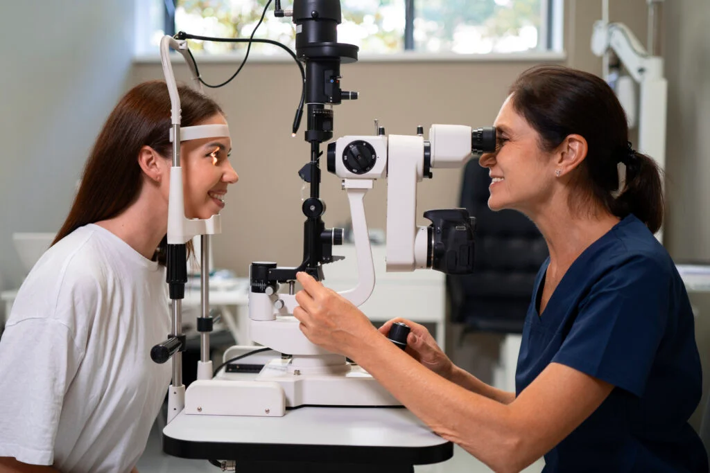
B-Scan ultrasonography is a non-invasive imaging technique used by ophthalmologists to assess the internal structures of the eye, including the retina. If you’re searching for the best retina specialist in Indore or the best eye hospital for RNFL scan in Indore, understanding how a B-Scan helps in diagnosing retinal detachment can be crucial for your eye health.
In this post, we’ll explore:
- What B-Scan ultrasonography is and how it works
- Why it is needed for diagnosing retinal detachment
- The benefits of B-Scan over other diagnostic methods
- Where to find the best eye retina specialist near you
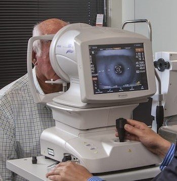
What is B-Scan Ultrasonography?
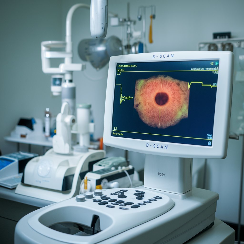
B-Scan (Brightness Scan) ultrasonography is an advanced imaging technique that uses high-frequency sound waves to create a cross-sectional image of the eye. It is particularly useful in cases where the retina cannot be directly visualized due to opacity in the eye’s media, such as in dense cataracts or vitreous hemorrhage.
How B-Scan Works
- A gel is applied to the closed eyelid.
- A small probe emits ultrasound waves that penetrate the eye.
- The sound waves bounce off different structures inside the eye and form an image.
- The ophthalmologist interprets the images to detect abnormalities like retinal detachment.
Why is B-Scan Needed for Retinal Detachment?
1. Detects Retinal Tears and Detachment
B-Scan is highly effective in identifying retinal detachment, especially in cases where the retina cannot be seen clearly with a traditional ophthalmoscope. Early detection can prevent permanent vision loss.
2. Useful in Cases of Opaque Media
If a patient has cataracts, vitreous hemorrhage, or corneal opacity, a standard eye exam may not provide a clear view of the retina. In such cases, B-Scan helps assess retinal health.
3. Helps in Post-Surgery Evaluation
After retinal surgeries, B-Scan can be used to check the success of the procedure and detect any complications like recurrent detachment or fluid accumulation.
4. Detects Other Eye Conditions
Apart from retinal detachment, B-Scan can help diagnose:
- Vitreous hemorrhage
- Intraocular tumors
- Optic nerve abnormalities
- Foreign bodies inside the eye
Comparison of B-Scan with Other Retinal Diagnostic Methods
| Diagnostic Test | Purpose | Best For | Limitations |
| B-Scan Ultrasonography | Uses ultrasound waves to create a cross-sectional image of the eye. | Retinal detachment, vitreous hemorrhage, tumors. | Cannot provide detailed retinal layer analysis. |
| Fundus Photography | Captures high-resolution images of the retina. | Monitoring retinal health over time. | Cannot see through cataracts or hemorrhage. |
| OCT (Optical Coherence Tomography) | Uses light waves to capture detailed images of the retina’s layers. | Retinal diseases like macular degeneration, diabetic retinopathy. | Cannot penetrate dense cataracts or vitreous opacities. |
| Fluorescein Angiography | Uses dye and a camera to study blood flow in the retina. | Detecting retinal vascular disorders. | Invasive, involves dye injection. |
Benefits of Getting a B-Scan at a Top Eye Hospital
If you’re looking for the best eye hospital for RNFL scan in Indore, you should consider a facility that offers comprehensive retinal imaging and diagnosis, including B-Scan. Some benefits of choosing a reputed eye retina specialist near you include:
- Advanced Diagnostic Equipment: Ensures accurate results and better treatment planning.
- Experienced Retina Specialists: Skilled in diagnosing and treating complex retinal conditions.
- Comprehensive Eye Care: Access to multiple eye tests like RNFL scans, OCT, and fundus photography in one place.
Where to Find the Best Retina Specialist in Indore?
If you are experiencing sudden vision loss, flashes of light, or floaters, you should immediately visit an expert retina specialist. The best eye hospital for RNFL scan in Indore will provide retina evaluations, including B-Scan ultrasonography.
When choosing an eye retina specialist near you, look for:
✔ A hospital with advanced diagnostic facilities.
✔ A specialist with expertise in retinal disorders.
✔ Positive patient reviews and high success rates in retinal treatments.
Schedule an appointment today with a leading eye retina specialist near you and ensure your eye health is in expert hands. 🚀
FAQs
1. How long does a B-Scan eye test take?
A B-Scan typically takes 5-10 minutes and is completely painless. The results are available immediately for interpretation by an ophthalmologist.
2. Is B-Scan safe for the eyes?
Yes, B-Scan uses ultrasound technology, which is non-invasive and completely safe for eye examinations.
3. Can B-Scan detect all retinal problems?
B-Scan is excellent for detecting retinal detachment, vitreous hemorrhage, and tumors, but it may not provide detailed retinal layer analysis like OCT scans.
Conclusion
B-Scan ultrasonography is a crucial tool for diagnosing retinal detachment and other retinal conditions, especially when the eye’s internal structures are not visible through standard exams. If you suspect a retinal issue, visit the best retina specialist in Indore for a thorough check-up.If you’re searching for the best eye hospital for RNFL scan in Indore, make sure they offer B-Scan, OCT, and other advanced retinal imaging services. Don’t delay an eye exam—early detection and timely treatment can protect your vision!